
胶囊内镜诊断出一名儿童的小肠多发性婴儿肌纤维瘤病
概要:
Infantile myofibromatosis (IMF) is a rare group of reactive lesions. Their clinical, radiological, and pathological features may be confused with those of malignancy. The lung and mediastinum are the most common site of IMF in all pediatric age groups .
We present a case of a 9-year-old boy presenting with a history of recurrent abdominal pain and chronic diarrhea since the age of one. He was under the third percentile for weight (16 kg) and height (106cm). Physical examination and laboratory findings were normal. Abdominal computed tomography (CT) showed a nodular lesion of the liver (segment IV), cholelithiasis, and nephrolithiasis. CT of the thorax showed a lesion measuring 3.9x2.2x2.6cm in the inferior left lung with bronchial and pulmonary vein invasion and mediastinal lymph nodes.
The patient underwent upper endoscopy and a subepithelial lesion with a central depression was found in the greater curvature of the gastric body. Colonoscopy showed multiple subepithelial lesions with a central ulceration, some of them with fibrin, varying in size from 3 mm to 20 mm ( Fig.1). Video capsule endos- copy(MiroCam; Intromedic, Seoul, Korea) showed two lesions in thejejunum similar to those found in the colon and stomach ( Fig.2; Video 1). Endoscopic ultrasonography of the colon with 15-MHz miniprobes showed a well-delimited, hypoechogenic, homogeneous, fusiform lesion with a central ulceration originating from the muscle layer, measuring 9.2×13.2mm ( Fig.3).
一名9岁男童从1岁开始就有周期性腹痛及隐性腹泻的病史。体格检查和实验室指标均正常,腹部CT显示肝部有结节状病变。患儿行上消化道内镜检查,在胃体胃大弯发现一处中心凹陷的上皮内病变。结肠镜检查发现多处中心溃疡的上皮内病变,大小从3mm到20mm不等。经MiroCam胶嚢内镜检查发现空肠有两处与胃部、结肠相似的病变。
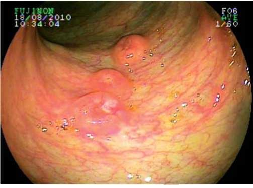
Fig.1 Colonoscopic image of the subepithelial lesion.
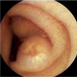
Fig 2 Video capsule endoscopy showing two subepithelial lesions in the jejunum.
Video capsule endoscopy shows small-bowel tumors in multicentric infantile myofibromatosis.
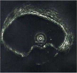
Fig.3 Endoscopic ultrasonography of the colon with 15-MHz miniprobes showing a well- delimited, hypoechogenic, homogeneous fusiform lesion with a central ulceration, originating from the muscle layer.
Biopsies were taken from the gastric and colonic lesions and a pathological diagnosis of IMF was made. The peripheral area showed spindle cells (myofibroblasts) with eosinophilic cytoplasm and ovoid nuclei, with less differentiated, rounder cells with pale cytoplasm and basophilic, small round nuclei in the central portion. No cellular anaplasia was present and there were few mitoses. The mesenchymal cells stained with vimentin and actin, but were uniformly negative with CD117, CD34, Ki-67, MIB-1, and S-100 antigen. Aerobic, anaerobic, and mycobacterial cultures obtained from the specimen were negative.
Endoscopy_UCTN_Code_CCL_1AC_2AC
Competing interests: None
L. R. Alberti1, P. F. Souto Bittencourt2,
A. Rodrigues Ferreira2,
R. R. Rodrigues Da Silva2,
S. Diniz Carvalho2,
F. M. B. Nunes Cachicolo2, M. Bahia2,
L. P. Fonseca de Castro2
1 Department of Surgery, Federal University of Minas Gerais, Santa Casa de beloHorizonte. Belo Horizonte. brazil
2 Department of Pediatrics, Federal University of Minas Gerais. Santa Casa de beloHorizonte. Belo horizonte. brazil
References
1 Lasso Betancor CE, Vazquez Rueda F, Vargas Cruz V et al. Congenital solitary infantile myofibroma: report of two cases. Cir Pediatr 2011;24:184-187
2 Smith A, Orchard D. Infantile myofibromato- sis: two families supporting autosomal dominant inheritance. Australas J Dermatol 2011; 52: 214-217
3 Al-Jazaeri A, Al-Zahem A, Al-Maziad H et al. Unilateral breast mass in an infant: a rare presentation of spontaneously regressing myofibromatosis. J Pediatr Surg 2010; 45: 1896-1899
4 Larralde M, Hoffner MV, Boggio P et al. Infantile myofibromatosis: report of nine patients. Pediatr Dermatol 2010; 27: 29 - 33


扫描二维码
关注华亘控股微信公众号
ICP经营许可证编号:京ICP备14021677号 北京华亘安邦科技有限公司 Design by CE ULITAME